
BIOMATERIALS LAB
Biomaterials Overview || Research Fields || Publications || PI Members-Alumni
Research Fields
Natural Biomaterials
As natural biomaterials, bone and silk-based materials are our main subjects of interest.
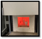
Bone is studied as a micro and nanostructured material with complex mechanical properties, demonstrating unique structure-function relations. We have studied the micro-to-nanostructure of bone using the environmental scanning electron microscope, ESEM and the ESPI. The mechanical properties of bone obtained using the ESEM and ESPI are modelled to understand the mineralized collagen structures on the mechanics of bone.

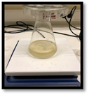
Ceramics and cement materials such as hydroxyapatite is another subject of concern for our lab. We have been working on the generation of synthetic hydroxyapatite using the Calcium and Phosphate containing materials and wet chemistry.
Synthetic Biomaterials
Ceramics
Ceramic materials and injectable cements are produced and characterized using standard techniques and instruments such as the SEM, XRD, FT-IR, Instron Universal Testing Device and Vicat Testing Device.
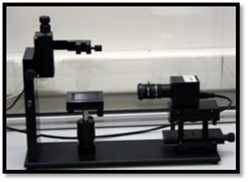
Materials are characterized using the contact angle measurements, using KSV CAM 101 contact angle measurement device (KSV Instruments Ltd., Finland) and 5 ml. of distilled water administered using Hamilton syringe. Twelve images are taken for each sample and recording started 5 seconds after the first drop. Images were taken with a 1 second interval for each specimen. Contact angle calculations were estimated using KSV CAM 101 contact angle program and average of the these intervals were taken as a final contact angle value.
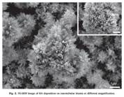
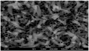
Surface morphology and elemental analysis of the prepared calcium phosphate materials are observed using scanning electron microscopy (Carl Zeiss, Germany). Porosity Estimation and Pore Distribution: ImageJ, an open source program, is used for SEM image processing to estimate porosity and pore distribution of the cement samples. Using the scale of the SEM image, 500 µm, is defined in pixels and used to measure the diameter of each pore the program toolbar (0.554 pixels/µm).
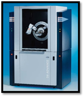
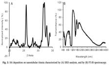
X-Ray diffraction is used to identify the elements and crystal structure of the synthesized and formulated and deposited calcium phosphate cements. XRD data (Bruker ASX, D8 ADVANCE, Berlin, Germany) was collected at 4° to 70° 2θ range.
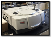
FTIR is used to determine bonding characterization of calcium phosphate cements using Nicolet IS50 ATR spectroscopy device (Madison, USA). Samples are crushed into fine powder using agate mortar and pestle and placed into the FT-IR device and data is collected using the OMNIC 9 software, where the experiment setup is adjusted to 16 number of scans with a resolution of 4 cm-1 in the wavenumber range of 400-4000 cm-1.
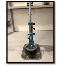
Setting time of a cement is the length of time required for the cement to solidify. Conventionally, Vicat apparatus is used to determine the setting time of cements.
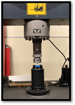
Materials are also compression tested using Instron Universal Testing Machine (Norwood, USA) giving a force-displacement curve, from which compressive strength calculations were obtained by normalising the load in Newtons with the cross sectional area of the specimen.
Surface Coatings
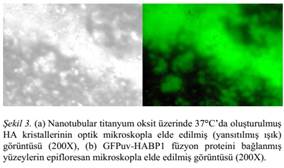
We are interested in Electrochemical Deposition of HA using the potentiostat. The application area of electrochemical deposition is in orthopedics and dentistry. This has been demonstrated with fusion proteins binding to HA coated surfaces.
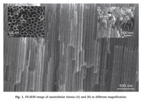
Coating of Orthopedic/Dental Implants using electrochemically deposited Hydroxyapatite requires the modification of Titanium Implants Surfaces. The cytotoxicity and surface reaction of Titanium implants with has been studied using modified Titanium and osteoblast.
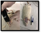
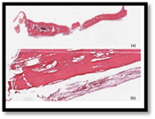
Animal experiments are conducted to investigate the osteoconductive and osteoinductive behavior of the Titanium implants, hydroxyapatite coated implants and injectable calcium phosphate cements in the bone tissue of a living organism; in this case, male 8-10 week old Sprague-Dawley rats.
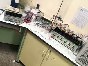
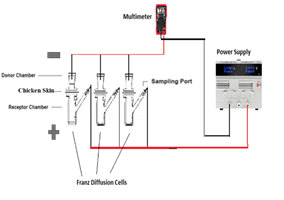
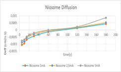
Iontophoresis Studies with Franz Diffusion Cell: Iontophoresis is the use of small electric currents to deliver the active substance to the body at a controlled rate. The electric current is carried by the drug ions and other ions in physiological liquids. Substance molecules can be electrically pushed or pulled through membranes quickly and controlled. The power supply was set to deliver the necessary amount of current to each sample each time which were 1mA, 2.5mA and 5mA respectively. As the experiment proceeded due to the change in system resistance the voltages were altered to maintain a stable current flow.
Charges and the sizes of the niosomes were measured with the use of Zetasizer. Samples collected from the sample port of the receptor chambers are examined under HPLC to determine the concentration of the drug that passed through the chicken skin over time.
High-performance liquid chromatography (HPLC) is used as an analytical technique to separate, identify, and quantify components in a mixture. A pump forces a solvent through a column under high pressures of up to 400 atmospheres. Instruments in the Zetasizer range are used to measure particle and molecular size from less than a nanometer to several microns using dynamic light scattering; zeta potential and electrophoretic mobility using electrophoretic light scattering; and molecular weight using static light scattering. Light scattering is a fundamental analytical technique for the characterization of particulate materials, and is most commonly applied to colloidal systems, nanoparticles and macromolecules in solution or dispersion, to determine particle size, molecular weight, or electrophoretic mobility. Different methods of light scattering analysis provide a range of useful information about your samples: Dynamic Light Scattering (DLS) measures the size and size distribution of molecules and particles and Electrophoretic Light Scattering (ELS) measures the electrophoretic mobility of particles or molecules in dispersion or solution – this is often converted to a ‘zeta potential’.
Biosensors is another area of interest that has a large application area in the field of sensors and actuators. We are interested in the use of affinity sensors in detection of cancer biomarkers as metabolites using small molecule detection.